Wide therapeutic window for melatonin in stroke
 Neuroprotective agents for stroke continue to fail in clinical trials. One important reason is that the therapeutic window for many of those agents is too narrow to confer benefits to acute stroke victims. It would be desirable to have a potent neuroprotectant agent that has a wide therapeutic window, few side effects, and can be easily obtained at low costs. Melatonin is a compound that seems to confer strong neuroprotective benefits in cerebral ischemia and is available in the United States as a dietary supplement.
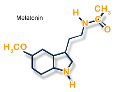
Melatonin is a potent antioxidant whose small molecular size and high lipophylicity makes for superior blood-brain barrier (BBB) permeability and penetration of the cell nucleus. Melatonin inhibits the production of inducible nitric oxide (iNOS), which exacerbates post-ischemic cell-death, and is a potent scavenger of peroxynitrite. The neuroprotective effects of melatonin have been observed in animal models of ischemic stroke when delivered after stroke, or as prophylaxis up to 9 weeks pre-stroke.
Additionally, melatonin exhibits very few side effects in humans. A previous (2005) meta-analysis of fourteen melatonin experiments documented an estimated improvement in outcome of 42.8%, though no studies used aged, hypertensive, or diabetic (well-known risk factors for stroke) animals, nor was melatonin administration investigated beyond 2 hours post-ischemia. However, In their recent article, Kilic et. al demonstrate that melatonin continues to provide significant neuroprotection even when administered 24 hours post-ischemia.
The researchers induced mild focal cerebral ischemia in a mouse model of middle cerebral artery (MCA) occlusion (30 minutes of MCA occlusion followed by reperfusion). They were then either left untreated, treated with vehicle only (ethanol in drinking water), or treated with melatonin (0.025 mg/mL) dissolved in ethanol in drinking water. Treatment was administered 24 hours after reperfusion via intraperitoneal bolus injection of 4mg/kg body weight (b.w.) melatonin, or via injection of the diluent in animals treated with vehicle. Continued treatment was administered through the animals’ drinking water at approximately 4 mg/kg b.w./day for 29 consecutive days. Two other groups of sham-operated animals were also either treated or untreated to serve as controls.
Prior to ischemic induction a baseline of grip strength, motor coordination, and spontaneous locomotor activity was measured in all animals. They were assessed again 7 and 30 days post-ischemia. Immunohistochemistry was then performed to characterize cell proliferation and neural progenitor cells.
The authors note that in untreated animals, 30 minutes of MCA occlusion resulted in “disseminated neuronal injury in the striatum, but not in the overlying cortex,” and that the percentage of surviving striatal neurons was significantly higher in ischemic animals treated with melatonin. This indicates a strong “structural rescue” effect of melatonin even when administered with a 24 hour delay. Melatonin also stimulated cell proliferation in the ischemic animals’ brains, evidenced by increased numbers of BrdU and DCX positive cells in animals treated with melatonin as compared to controls.
Melatonin exhibits a beneficial psychomotor effect in post-ischemic animals. Both grip strength and motor coordination measures were significantly higher in melatonin treated animals — in fact, motor coordination was improved almost to nonischemic animals’ level by 7 days, an effect which persisted at 30 days. In contrast, animals treated only with vehicle exhibited severe deficiencies in grip strength and motor coordination at 7 and 30 days post-ischemia.
Post-ischemic anxiety and hyperactivity were also attenuated in melatonin treated animals as measured by open-field observations of spontaneous locomotor activity. Because post-ischemic anxiety in animals is hypothesized to be analagous to post-ischemic depression in humans, these beneficial psychomotor effects could be especially advantageous in ameliorating the psychopathological symptoms associated with stroke.
These results indicate a wide therapeutic window for melatonin administration to reduce brain injury after stroke. Melatonin has now been shown to demonstrate potent neuroprotective effects when given before, immediately after, and up to 24 hours after stroke. Melatonin is also a core component in an anti-oxidant “cocktail,” developed by Michael Darwin et al., called VitalOxy, to mitigate reperfusion injury after cardiac arrest in cryonics patients. But its greater potential lies in prophylactic use. For cryonics patients, premedication of melatonin in the days or weeks leading up to pronouncement of medico-legal death can be a real, practical, and easy way to ensure appropriate “pre-mortem” protection against brain injury after legal pronouncement of death.
In their review article, Reiter et al. (2005) point out that melatonin not only limits cellular destruction of the brain due to ischemia, but also of other tissues, such as the heart. Melatonin’s ability to significantly improve outcome in stroke warrants serious consideration of melatonin as a neuroprotectant in everyday medical settings. Because melatonin is an endogenous molecule (i.e., it is produced by the body naturally), it is unpatentable so pharmaceutical companies do not stand to make much of a profit from the widespread use of melatonin, which severely limits the promotion of melatonin for use in stroke treatment.
Melatonin is widely available and inexpensive, making it an attractive option for both stroke patients and candidates for human cryopreservation. However, an advisable human dosage for cerebroprotection is difficult to determine — although the side effects of melatonin are minimal, it is well known that melatonin causes lethargy and induces sleep in most humans at much lower dosages than those described in the experimental literature. Unfortunately, the relevant research articles leave us clueless as to the general physiological effects of high doses of melatonin: the animals’ sleeping patterns or activity levels are not mentioned.
Improved protection of the brain after stroke may be achieved when melatonin is combined with other dietary supplements that have a wide therapeutic window and complement the action of melatonin by mitigating other elements in the ischemic cascade such as supporting energy generation in the brain, inhibition of PARP, and inhibition of apoptosis.